



Indigenous Placenta-on-Chip Transform Platform
| General Studies Paper III: Biotechnology, Health |
Why in News?
Recently, researchers from IIT Bombay and ICMR-National Institute for Research on Women’s Health (NIRWoH) developed an indigenous placenta-on-chip platform.
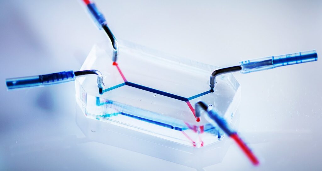
Image Credit: Phys.org
What is the Indigenous Placenta-on-Chip Platform?
- About: Placenta-on-chip is a microfluidic bioengineered devicethat recreates the human placental barrier using living cells.
- It enables scientists to study nutrient transfer, drug transport, and disease mechanisms during pregnancy.
- Indian researchers recently developed an indigenous version, reducing dependence on imported platforms.
- Developers: The platform was developed by IIT Bombay in collaboration with ICMR-National Institute for Research on Women’s Health.
- The work was reported in 2025 and represents a major step in India’s biomedical and organ-on-chip research ecosystem.
- Technology Used: The device uses microfluidic channels that mimic maternal and fetal blood flow.
- The devices were built using photolithography and soft lithography.
- Human placental cells are cultured on a membrane, creating a dynamic 3D environment unlike conventional static petri-dish experiments.
- Placental Functions Simulated: The chip reproduces key placental functions such as oxygen exchange, nutrient transport, waste removal, and hormonal interactions.
- This allows researchers to observe how substances move between mother and fetus.
- Medical Importance: The platform can help investigate pregnancy disorders such as preeclampsia, gestational diabetes, and fetal growth restriction, which are major contributors to maternal and neonatal mortality in India.
- Drug Testing: Researchers can test whether medicines, nanoparticles, or environmental toxins cross the placental barrier.
- This is crucial because pregnant women are often excluded from clinical trials, creating a major knowledge gap in drug safety.
- Animal Model Advantage: Animal placentas differ significantly from human placentas. The chip offers human-specific data, potentially improving prediction accuracy and reducing the need for animal experimentation.
- Research Applications: Potential applications include personalized medicine, toxicology studies, vaccine safety assessment, nutrition research, and AI-driven biomedical modeling.
- Real-Time: The completed chip enables real-time observation of maternal–fetal interactions, cellular responses, transport kinetics, and endocrine activity under controlled experimental conditions.
- Support: The project received support from IMPRINT II C of the Science, Engineering, and Research Board, or SERB.
- The patent filing for this technology is under process.
How Does Placenta-on-Chip Technology Work?
- The platform recreates the maternal–fetal interface using a two-compartment microphysiological system.
- Separate maternal and fetal chambers reproduce the spatial organization of the human placenta, enabling realistic laboratory simulation of placental transport processes.
- Both chambers are separated by a microporous semipermeable membrane coated with extracellular matrix (ECM) proteins.
- This membrane functions like the natural placental barrier, permitting selective molecular exchange.
- Scientists culture human trophoblast cells on the maternal side and human endothelial cells on the fetal side.
- These cells organize into a bilayer barrier, closely resembling the structural arrangement found in a healthy human placenta.
- The trophoblast cells are induced to undergo syncytialisation, forming a syncytiotrophoblast-like layer.
- This mature tissue secretes β-human chorionic gonadotropin (β-hCG), confirming tissue behaves similarly to a functional placenta.
- Researchers introduce nutrients, drugs, or test compounds into the maternal chamber and monitor their controlled diffusion across the engineered barrier.
- Transport occurs only when permitted by the biological barrier, replicating physiological selectivity.
- The platform continuously measures glucose flux, urea clearance, permeability, and molecular movement between compartments.
- These quantitative data reveal transport efficiency, barrier integrity, and metabolic performance under laboratory conditions.
- Researchers can recreate hyperglycaemic conditions resembling gestational diabetes by altering culture media composition.
- The platform then records changes in transport dynamics without disrupting barrier integrity, enabling disease modelling.
- Barrier performance is validated by analysing selective permeability, cellular junction formation, and transport restriction.
- Macromolecules remain largely blocked, whereas essential small molecules cross under regulated conditions.
Points to Know
- The placenta is a temporary fetomaternal organ that develops inside the uterus during pregnancy.
- It connects the developing fetus to the mother’s uterus through the umbilical cord, acting as the primary life-support system until birth.
- Placental development begins about 6–7 days after fertilization, when the blastocyst implants into the uterine lining.
- It develops throughout pregnancy, with rapid formation during the first trimester, while major maturation continues until delivery at approximately 40 weeks.
- The placenta consists of a fetal component (chorion frondosum) and a maternal component (decidua basalis).
- Millions of microscopic chorionic villi greatly increase the surface area for efficient exchange between maternal and fetal circulations.
- A mature placenta is disc-shaped, measuring approximately 20–22 cm in diameter, 2–3 cm thick, and weighing about 450–500 g at full term.
- The umbilical cord inserts into its fetal surface, carrying blood between the fetus and placenta.
- Its principal function is the bidirectional exchange of oxygen, glucose, amino acids, fatty acids, vitamins, water, and electrolytes from mother to fetus while removing carbon dioxide, urea, and metabolic wastes from fetal circulation without direct blood mixing.
- The placenta functions as a major endocrine organ, producing hormones including human chorionic gonadotropin (hCG), progesterone, estrogens, human placental lactogen (hPL), placental growth factor (PlGF), and corticotropin-releasing hormone (CRH).
- The placental barrier limits the passage of many pathogens while allowing transfer of maternal IgG antibodies, providing passive immunity to the fetus.
- Following childbirth, the placenta is expelled during the third stage of labour (afterbirth), usually within 30 minutes.
FAQs:
- What is the Placenta-on-Chip platform?
A microphysiological device that replicates the human placental barrier for studying maternal–fetal interactions in the laboratory. - Who developed the Placenta-on-Chip platform in India?
Researchers from IIT Bombay and ICMR–National Institute for Research on Women’s Health (NIRWoH) jointly developed the indigenous platform. - How does the Placenta-on-Chip technology work?
It uses human placental and endothelial cells on a porous membrane to simulate maternal–fetal transport and barrier functions. - Why is the Placenta-on-Chip platform important for pregnancy research?
It enables safe study of placental biology, fetal development, and pregnancy disorders under controlled laboratory conditions. - How can this technology improve drug testing?
It predicts drug transfer across the placenta, improving safety assessment of medicines intended for pregnant women. - What are the advantages of organ-on-chip technology?
It provides human-relevant, reproducible, cost-effective models with better physiological accuracy than conventional cell cultures. - Can the Placenta-on-Chip reduce animal testing?
Yes. It offers a human-based alternative, reducing reliance on animal experiments for placental and drug research. - What medical conditions can be studied using this platform?
It can study preeclampsia, gestational diabetes, placental dysfunction, and impaired maternal–fetal transport.
Disclaimer: Information in this article is based on official announcements and public records. Regulations and implementation details may evolve over time.